Bioflow Considerations in Cervical Spine Related Headache
Alex Nichols, FRS Instructor
Numerous research papers have mentioned the connection between some of the deeper tissues of the cervical spine like the RCPM and RCPm being continuous with the dura mater. This is clinically significant because (A) this is further evidence for the Bioflow model of anatomy and physiology, (B) in FR we learn that being able to locate, assess, and treat such tissues are some of the objective goals manual therapy should be encompassing but currently is not, and finally, (C) we can see how these deeper tissues of the cervical spine would be important tissues to assess in client complaints around headaches or related complaints when looking at clinical history and intake.
Let’s briefly review the anatomy of the structures to be discussed.
The rectus captious posterior major (RCPM) – shares innervation with RCMm via the suboccipital nerve, originating from this spinous process of C2 and inserting onto the inferior nuchal line of the occiput.
Rectus capitous posterior minor (RCPm) is innervated by the suboccipital nerve, originating from the posterior arch of C1 and inserting onto the occiput inferior to the nuchal line.
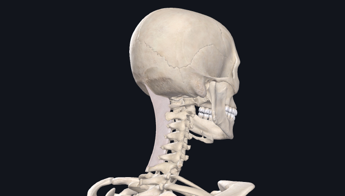
The Nuchal Ligament is a large fibre-elastic connective tissue based structure that extends from the EOP (external occipital protuberance) down through the spinous processes of the cervical vertebrae through C7. Because it is a tissue that spans deep to superficial and has a large surface area to access, this makes it much easier from a palpation standpoint to access versus deeper tissues like RCPM and RCPm where we would need to layer through other tissues of the cervical spine.
 Fig 1. Right Suboccipitals (posterior view)
Fig 2. Nuchal Ligament (postero-lateral view)
The clinical significance of these tissues in relation to the Bioflow model is, “A connective tissue link between the rectus capitis posterior minor (RCPMi) muscle and the cervical dura mater, hereafter referred to as the ‘‘myodural bridge’’ as originally described by Hack et al. (1995). It’s been implicated as a source of cervicogenic headache pain. Subsequent investigators have verified this connection and described attachments between the cervical dura mater and nuchal ligament (Mitchell et al., 1998; Dean and Mitchell, 2002; Humphreys et al., 2003). There is now solid evidence for the existence of this structure and its clinical importance in relation to cervicogenic headache. This readily dissected anatomical feature has not yet been presented in any of the commonly used medical anatomy texts.
What does this mean from an intervention standpoint? As FR therapists, we understand that tissues innervated by spindles will have two outcome measures possible whereas those that lack innervation are largely connective tissue based and will have only one outcome measure. The length/tension capacities in individual tissues contributing to the overall ability, or lack thereof, to express joint motion at a level such as the suboccipitals or nuchal ligament as they blend into capsular and subsequently dura mater attachments. We therefore can have a direct impact on problematic findings in such areas due to their connection. Moreover, global or local tissue-based findings are not sufficient for proper functioning of such tissues and would be associated with fibrotic depositions and/or atrophy.
“Evidence exists that dysfunction of the RCPMi is involved in the etiology of headache. Elliott et al. (2005, 2006, 2008) demonstrated through a series of MRI studies that the RCPMi of individuals with chronic neck pain due to whiplash trauma contained significant amounts of fatty infiltration, possibly due to damage of the suboccipital musculature.“
Further, there is evidence to suggest that greater atrophy of these tissues specifically has been shown to be associated with grater headache intensity, duration, or frequency. Whether or not this is a primary cause, we can’t ignore the overall importance of what seems to occur when such tissues atrophy due to injury or other pathologies. As such, understanding the available real estate we can access from a manual therapist perspective, how to get to such levels and specifically intervene using FR principles cannot be understated. If the client or patient presents with problems like creating length in these areas or presents with aberrant tension due to fibrosis or atrophy, understanding how these tissues fit into the bioflow model and have an effect on neck function at large is our responsibility.
References:
Fernández-de-las-Peñas, C., Bueno, A., Ferrando, J., Elliott, J., Cuadrado, M., & Pareja, J. (2007). Magnetic Resonance Imaging Study of The Morphometry of Cervical Extensor Muscles in Chronic Tension-Type Headache. Cephalalgia, 27(4), 355–362.doi:10.1111/j.1468-2982.2007.01293.x
