The Compressor Cuff
By: Dr. Mitch Broser, FRS Instructor @MBroser
The Rotator Cuff is made up of 4 muscles: subscapularis, supraspinatus, infraspinatus and teres minor. With a better understanding of their function, more recent literature refers to these structures as the “Compressor Cuff”. These four muscles originate from the scapula and contribute their distal tendinous fibers to the glenohumeral joint capsule.
The subscapularis is the largest of the four muscles, attaching distally to the lesser tubercle of the humerus. It’s distal fibers contribute to the anterior glenohumeral joint capsule and transverse humeral ligament bioflow, where the long head of biceps tendon is held within the intertubercular groove. From anterior to posterior, the supraspinatus, infraspinatus and teres minor attach onto the greater tubercle of the humeral as they become the glenohumeral joint capsule.
The cuff footprint refers to the humeral attachment sites of the cuff tendons. Cadaveric studies have proven this to be an inadequate way to describe these distal insertions. More accurately, the distal tendons and glenohumeral joint capsule are one complete, inseparable complex. The fibers of the cuff tendons interdigitate not only with one another, but also fuse together with the internal joint capsule along its deepest layer before the common insertion into bone.
The supraspinatus is made up of an anterior and posterior muscular belly. The anterior belly is much larger, while its tendon is much thicker and more tubular compared to the smaller posterior belly and its broad, flat tendon. The anterior musculotendinous unit can experience up to 2.88 times as much load as the posterior portion, often contributing to unequal force distribution across the distal tendon (Roh, et al. 1999). The thicker anterior edge of the supraspinatus tendon forms a “stress riser zone” between the anterior and mid-posterior fibers. This may help explain why more than 90% of degenerative tears start and propagate from this junction (Pandey et al. 2015). Differential tissue organization between various layers within or between cuff tendons initiates stress risers and can cause intrasubstance tears, which are twice as common as bursal- or articular-side tears. Stress-failure point of the articular side is half of bursal side because robust collagen on the bursal side makes is more resistant to tensile stress.
Some less talked-about anatomy of the cuff is the rotator cable, rotator interval and rotator crescent. The rotator cable (aka. The semicircular ligament of the humerus, or circular fiber system) is a curved extension of the coracohumeral ligament which stretches between the lesser tuberosity and the inferior facet of the greater tuberosity (ie. from subscapularis to teres minor). The cable runs in an anterior to posterior direction (perpendicular to the cuff tendons), underneath the supraspinatus and infraspinatus tendon, with a primary function of distributing forces across the cuff complex. The cable provides a stress-shielding effect to the thinner rotator crescent tissue, located just distal, by distributing forces across the span of the humeral head.
The rotator crescent is a thin crescent-shaped sheet of the rotator cuff, consistent with the lateral-most end of supraspinatus and infraspinatus tendinous sheet. The Rotator Crescent is an avascular zone which tears easily. Older individuals are typically cable dominant (thicker cable compared to crescent), whereas younger individuals are crescent-dominant (thicker crescent compared to cable).
(Asghar, 2020)
There are two Rotator Intervals (RI) – anterior and posterior. The anterior RI is in the anterosuperior part of the shoulder joint, with borders formed by the supraspinatus, subscapularis and the base of the coracoid process. The anterior RI includes the extra-articular coracohumeral ligament (CHL), the superior and middle glenohumeral ligaments, long head biceps tendon and glenohumeral capsule. The posterior interval is between supraspinatus and infraspinatus. The overall function of the RI is to (1) act as a shackle to prevent extreme flexion, extension, adduction and external rotation; (2) stabilize the humeral head against inferior translation during extreme adduction; (3) steady the humeral head against posterior translation during extreme flexion or external rotation of abducted shoulder (Asghar, 2020).
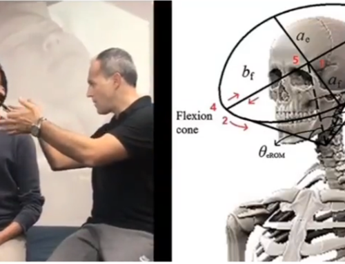
The primary function of the cuff complex is to keep the head of the humerus depressed and centered in the glenoid fossa permitting a focused center of rotation. When someone experiences tissue pathology of the cuff, the cuff cannot maintain the humeral head depressed and centered in the glenoid fossa. With accessory humeral head movement, variability of shoulder joint motion is reduced as the shoulder can only explore a limited area of capsular workspace. This accessory movement often presents with aberrant joint function, or a closing angle finding) in the shoulder.
When someone presents with a closing angle finding, we need to assess the deepest capsular tissue and intervene accordingly. For the shoulder, this tissue is the cuff. Using rotational based inputs, we can preferentially load the deepest tissue, particularly the capsular tissue and rotator cable. However, rotational-based inputs may not be as directionally specific for cuff tendons and humeral ligaments. With our understanding of how interconnected the cuff tendons, ligaments and capsular tissues are, and how tissue organization differs between these structures, we can approach shoulder capsule loading more comprehensively.
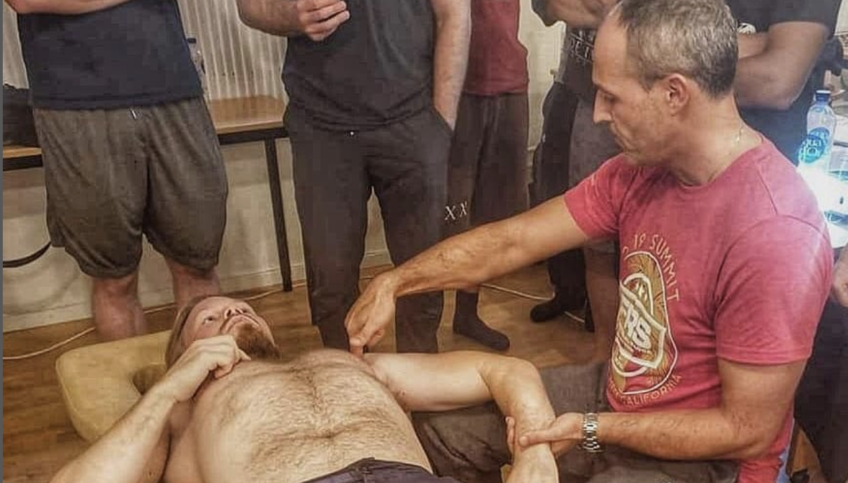
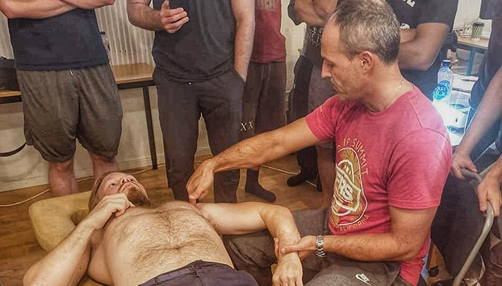
In FR Upper limb, we discuss FP® and FR® of the distal cuff tendons and multiple areas of the glenohumeral joint capsule. In cases of aberrant shoulder joint function, any of these tissues may present with poor tissue quality; what we refer to as mechanical tension. Normalizing these tissues with specific directional loading is needed. The tendinous and ligamentous structure run perpendicular to the capsule tissue, so load-based interventions should reflect that. We can preferentially load joint capsule or rotator cable tissues with rotational-based inputs, or tendinous tissues with more linear-based inputs.
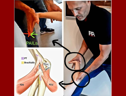
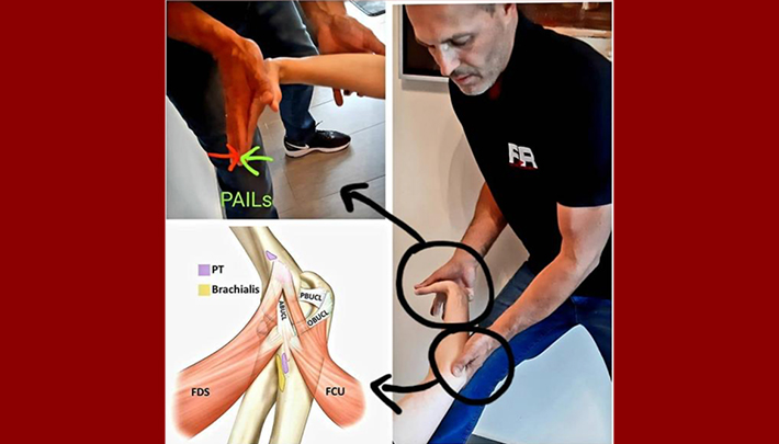
In FR, we use level 1 PAILs to begin the process of loading more bioflow with more force. As we progress into level 2 and eventually level 3 PAILs, we can continue to increase load magnitude to load more bioflow tissue. When managing the cuff, we need to progressively load with both rotational and linear inputs to improve tissue quality in all aspects of the capsular bioflow of the shoulder joint.
References:
1Asghar, A., Sanjib, KH., Ravi KN. Revisiting the anatomy of rotator cuff relevant to rotator cuff injury. National Journal of clinical anatomy. Vol 9. No 1. (2020).
2Pandey V., Willems WJ. Rotator cuff tear: a detailed update. Asia-pacific journal of sports medicine, arthroscopy, rehabilitation and technology 2. P1-14 (2015).
3Podgorski, MT., Loewnik, L., Grzelak., Polguj, M., Topol, M. Rotator cable in pathological shoulders: comparison with normal anatomy in a cadaver study. Anatomical science international. 94:53-57 (2019)
4Roh MS. et al. Anterior and posterior musculotendinous anatomy of the supraspinatus. 45th annual meeting, orthopaedic research society, Feb 1-4 (1999).

